
Tissues
In biology, tissue is a cellular organizational level between cells and a complete organ. A tissue is an ensemble of similar cells and their extracellular matrix from the same origin that together carry out a specific function. Organs are then formed by the functional grouping together of multiple tissues.
The English word is derived from the French tissu, meaning something that is woven, from the verb tisser, “to weave”.
The study of human and animal tissues is known as histology or, in connection with disease, histopathology. For plants, the discipline is called plant anatomy. The classical tools for studying tissues are the paraffin block in which tissue is embedded and then sectioned, the histological stain, and the optical microscope. In the last couple of decades, developments in electron microscopy, immunofluorescence, and the use of frozen tissue sections have enhanced the detail that can be observed in tissues. With these tools, the classical appearances of tissues can be examined in health and disease, enabling considerable refinement of medical diagnosis and prognosis.

Plant tissues
In plant anatomy, tissues are categorized broadly into three tissue systems: the epidermis, the ground tissue, and the vascular tissue.
- Epidermis – Cells forming the outer surface of the leaves and of the young plant body.
- Vascular tissue – The primary components of vascular tissue are the xylem and phloem. These transport fluid and nutrients internally.
- Ground tissue – Ground tissue is less differentiated than other tissues. Ground tissue manufactures nutrients by photosynthesis and stores reserve nutrients.
Plant tissues can also be divided differently into two types:
- Meristematic tissues
- Permanent tissues.
Meristematic tissues
Meristematic tissue consists of actively dividing cells, and leads to increase in length and thickness of the plant. The primary growth of a plant occurs only in certain, specific regions, such as in the tips of stems or roots. It is in these regions that meristematic tissue is present. Cells in these tissues are roughly spherical or polyhedral, to rectangular in shape, and have thin cell walls. New cells produced by meristem are initially those of meristem itself, but as the new cells grow and mature, their characteristics slowly change and they become differentiated as components of the region of occurrence of meristimatic tissues, they are classified as:
- Apical Meristem – It is present at the growing tips of stems and roots and increases the length of the stem and root. They form growing parts at the apices of roots and stems and are responsible for increase in length, also called primary growth. This meristem is responsible for the linear growth of an organ.
- Lateral Meristem – This meristem consist of cells which mainly divide in one plane and cause the organ to increase in diameter and growth. Lateral meristem usually occurs beneath the bark of the tree in the form of Cork Cambium and in vascular bundles of dicots in the form of vascular cambium. The activity of this cambium results in the formation of secondary growth.
- Intercalary Meristem – This meristem is located in between permanent tissues. It is usually present at the base of node, inter node and on leaf base. They are responsible for growth in length of the plant and increasing the size of the internode, They result in branch formation and growth.
The cells of meristematic tissues are similar in structure and have thin and elastic primary cell wall made up of cellulose. They are compactly arranged without inter-cellular spaces between them. Each cell contains a dense cytoplasm and a prominent nucleus. Dense protoplasm of meristematic cells contains very few vacuoles. Normally the meristematic cells are oval, polygonal or rectangular in shape.
Meristemetic tissue cells have a large nucleus with small or no vacuoles, they have no inter cellular spaces.

Permanent tissues
The meristematic tissues that take up a specific role lose the ability to divide. This process of taking up a permanent shape, size and a function is called cellular differentiation. Cells of meristematic tissue differentiate to form different types of permanent tissue. There are 3 types of permanent tissues:
- simple permanent tissues
- complex permanent tissues
- special or secretory tissues (glandular).
Simple tissues
A group of cells which are similar in origin; similar in structure and similar in function are called simple permanent tissue. They are of four types:

Parenchyma
Parenchyma (para – ‘beside’; chyma – ‘in filling, loose, unpacked’) is the bulk of a substance. In plants, it consists of relatively unspecialised living cells with thin cell walls that are usually loosely packed so that intercellular spaces are found between cells of this tissue. This tissue provides support to plants and also stores food. In some situations, a parenchyma contains chlorophyll and performs photosynthesis, in which case it is called a chlorenchyma. In aquatic plants, large air cavities are present in parenchyma to give support to them to float on water. Such a parenchyma type is called aerenchyma.
Collenchyma
Collenchyma is Greek word where “Collen” means gum and “chyma” means infusion. It is a living tissue of primary body like Parenchyma. Cells are thin-walled but possess thickening of cellulose, water and pectin substances (pectocellulose) at the corners where number of cells join together. This tissue gives a tensile strength to the plant and the cells are compactly arranged and have very little inter-cellular spaces. It occurs chiefly in hypodermis of stems and leaves. It is absent in monocots and in roots.
Collenchymatous tissue acts as a supporting tissue in stems of young plants. It provides mechanical support, elasticity, and tensile strength to the plant body. It helps in manufacturing sugar and storing it as starch. It is present in the margin of leaves and resist tearing effect of the wind.
Sclerenchyma
Sclerenchyma is Greek word where “Sclrenes” means hard and “chyma” means infusion. This tissue consists of thick-walled, dead cells. These cells have hard and extremely thick secondary walls due to uniform distribution of lignin. Lignin deposition is so thick that the cell walls become strong, rigid and impermeable to water.
Epidermis
The entire surface of the plant consists of a single layer of cells called epidermis or surface tissue. The entire surface of the plant has this outer layer of epidermis. Hence it is also called surface tissue. Most of the epidermal cells are relatively flat. The outer and lateral walls of the cell are often thicker than the inner walls. The cells forms a continuous sheet without inter cellular spaces. It protects all parts of the plant.
Complex permanent tissue
The complex tissue consists of more than one type of cells which work together as a unit. Complex tissues help in the transportation of organic material, water and minerals up and down the plants. That is why it is also known as conducting and vascular tissue. The common types of complex permanent tissue are:
- Xylem or wood
- Phloem or bast.
Xylem and phloem together form vascular bundles.
Xylem
Xylem consists of:
- Tracheid
- Vessel members
- Xylem fibers
- Xylem parenchyma
Xylem serves as a chief conducting tissue of vascular plants. It is responsible for the conduction of water and mineral ions/salt.
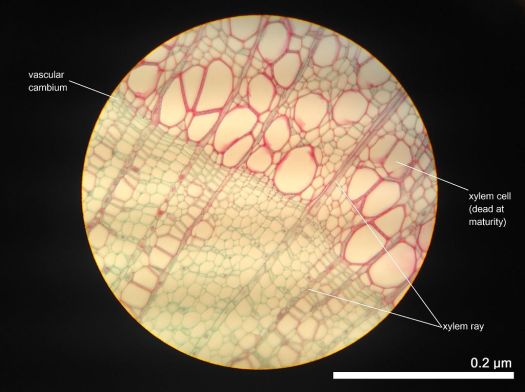
Cross section of 2 year old Tilia Americana, highlighting xylem ray shape and orientation
Xylem tissue is organized in a tube-like fashion along the main axes of stems and roots. It consists of a combination of parenchyma cells, fibers, vessels, tracheids, and ray cells. Longer tubes made up of individual cells are vessels (tracheae), while vessel members are open at each end. Internally, there may be bars of wall material extending across the open space. These cells are joined end to end to form long tubes. Vessel members and tracheids are dead at maturity. Tracheids have thick secondary cell walls and are tapered at the ends. They do not have end openings such as the vessels. The tracheids ends overlap with each other, with pairs of pits present. The pit pairs allow water to pass from cell to cell.
Though most conduction in xylem tissue is vertical, lateral conduction along the diameter of a stem is facilitated via rays. Rays are horizontal rows of long-living parenchyma cells that arise out of the vascular cambium. In trees and other woody plants, rays radiate out from the center of stems and roots, and appear like spokes on a wheel in cross section. Rays, unlike vessel members and tracheids, are alive at functional maturity.
Phloem
Phloem consists of:
- Sieve tube
- Companion cell
- Phloem fiber
- Phloem parenchyma.
Phloem is an equally important plant tissue as it also is part of the ‘plumbing system’ of a plant. Primarily, phloem carries dissolved food substances throughout the plant. This conduction system is composed of sieve-tube member and companion cells, that are without secondary walls. The parent cells of the vascular cambium produce both xylem and phloem. This usually also includes fibers, parenchyma and ray cells. Sieve tubes are formed from sieve-tube members laid end to end. The end walls, unlike vessel members in xylem, do not have openings. The end walls, however, are full of small pores where cytoplasm extends from cell to cell. These porous connections are called sieve plates. In spite of the fact that their cytoplasm is actively involved in the conduction of food materials, sieve-tube members do not have nuclei at maturity. It is the companion cells that are nestled between sieve-tube members that function in some manner bringing about the conduction of food. Sieve-tube members that are alive contain a polymer called callose, a carbohydrate polymer, forming the callus pad/callus, the colourless substance that covers the sieve plate. Callose stays in solution as long as the cell contents are under pressure. Phloem transports food and materials in plants upwards and downwards as required.
