
Plant anatomy or phytotomy is the general term for the study of the internal structure of plants. Originally it included plant morphology, the description of the physical form and external structure of plants, but since the mid-20th century plant anatomy has been considered a separate field referring only to internal plant structure. Plant anatomy is now frequently investigated at the cellular level, and often involves the sectioning of tissues and microscopy.
Structural divisions
Although some plant anatomy studies utilize a systems approach, such as the study of vascular tissues, plant anatomy is more classically divided into the following structural categories:
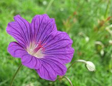
- Calyx-A sepal is a part of the flower of angiosperms (flowering plants). Usually green, sepals typically function as protection for the flower in bud, and often as support for the petals when in bloom.The term sepalum was coined by Noël Martin Joseph de Necker in 1790, and derived from the Greek σκεπη (skepi), a covering.
- Corolla-The role of the corolla in plant evolution has been studied extensively since Charles Darwin postulated a theory of the origin of elongated corollae and corolla tubes.A corolla of separate tepals is apopetalous. If the petals are free from one another in the corolla, the plant is polypetalous or choripetalous; while if the petals are at least partially fused together, it is gamopetalous or sympetalous. In the case of fused tepals, the term is syntepalous. The corolla in some plants forms a tube.
Androecium-The stamen (plural stamina or stamens) is the pollen-producing reproductive organ of a flower. Collectively the stamens form the androecium.
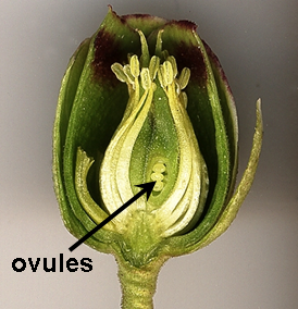
- Gynoecium-Gynoecium (from Ancient Greek γυνή, gyne, meaning woman, and οἶκος, oikos, meaning house) is most commonly used as a collective term for the parts of a flower that produce ovules and ultimately develop into the fruit and seeds. The gynoecium is the innermost whorl of (one or more) pistils in a flower and is typically surrounded by the pollen-producing reproductive organs, the stamens, collectively called the androecium. The gynoecium is often referred to as the “female” portion of the flower, although rather than directly producing female gametes (i.e. egg cells), the gynoecium produces megaspores, each of which develops into a female gametophyte which then produces egg cells.
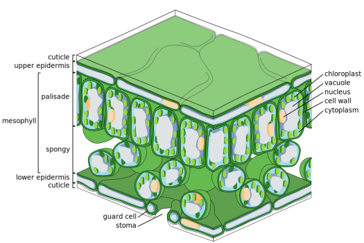
- Epidermis-The epidermis (from the Greek ἐπιδερμίς, meaning “over-skin”) is a single layer of cells that covers the leaves, flowers, roots and stems of plants. It forms a boundary between the plant and the external environment. The epidermis serves several functions: it protects against water loss, regulates gas exchange, secretes metabolic compounds, and (especially in roots) absorbs water and mineral nutrients. The epidermis of most leaves shows dorsoventral anatomy: the upper (adaxial) and lower (abaxial) surfaces have somewhat different construction and may serve different functions. Woody stems and some other stem structures[which?] produce a secondary covering called the periderm that replaces the epidermis as the protective covering.
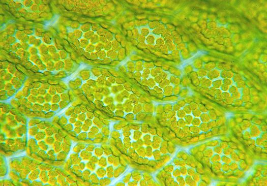
- Palisade cells-Palisade cells are plant cells loca]] in leaves, right below the epidermis and cuticle. They are vertically elongated, a different shape from the spongy mesophyll cells beneath them in the leaf. Their chloroplasts absorb a major portion of the light energy used by the leaf. Palisade cells occur in dicotyledonous plants, and also in the net-veined monocots, the Araceae and Dioscoreaceae.
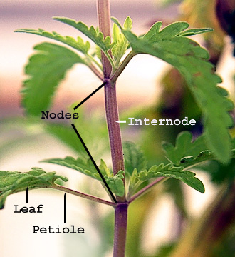
- Stem anatomy
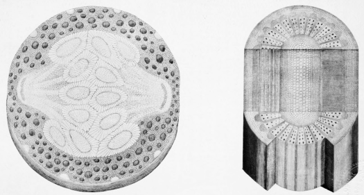
- Stem structure-Stem usually consist of three tissues, dermal tissue, ground tissue and vascular tissue. The dermal tissue covers the outer surface of the stem and usually functions to waterproof, protect and control gas exchange. The ground tissue usually consists mainly of parenchyma cells and fills in around the vascular tissue. It sometimes functions in photosynthesis. Vascular tissue provides long distance transport and structural support. Most or all ground tissue may be lost in woody stems. The dermal tissue of aquatic plants stems may lack the waterproofing found in aerial stems. The arrangement of the vascular tissues varies widely among plant species.
- Fruit/Seed anatomy
- Ovule-In seed plants, the ovule (“small egg“) is the structure that gives rise to and contains the female reproductive cells. It consists of three parts: The integument(s) form its outer layer(s), the nucellus (or remnant of the megasporangium), and female gametophyte (formed from haploid megaspore) in its center. The female gametophyte—specifically termed a megagametophyte—is also called the embryo sac in angiosperms. The megagametophyte produces an egg cell (or several in some groups) for the purpose of fertilization
- .Seed structure-A seed is an embryonic plant enclosed in a protective outer covering. The formation of the seed is part of the process of reproduction in seed plants, the spermatophytes, including the gymnosperm and angiosperm plants.Seeds are the product of the ripened ovule, after fertilization by pollen and some growth within the mother plant. The embryo is developed from the zygote and the seed coat from the integuments of the ovule.
- Pericarp-Fruits are the mature ovary or ovaries of one or more flowers. In fleshy fruits, the outer layer (which is often edible) is the pericarp, which is the tissue that develops from the ovary wall of the flower and surrounds the seeds.But in some seemingly pericarp fruits, the edible portion is not derived from the ovary. For example, in the fruit of the ackee tree the edible portion is an aril, and in the pineapple several tissues from the flower and stem are involved.
Accessory fruit-An accessory fruit (sometimes called false fruit, spurious fruit, pseudofruit, or pseudocarp) is a fruit in which some of the flesh is derived not from the ovary but from some adjacent tissue exterior to the carpel. Examples of accessory tissue are the receptacle of the strawberry, pineapple, common fig, and mulberry, and the calyx of Gaultheria procumbens or Syzygium jambos. Pomes, such as apples and pears, are also accessory fruits, with much of the fruit flesh derived from a hypanthium. Other example could be the anthocarps specific to the family Nyctaginaceae, where most of the fruit comes from the perianth (floral whorls).
- Wood anatomy
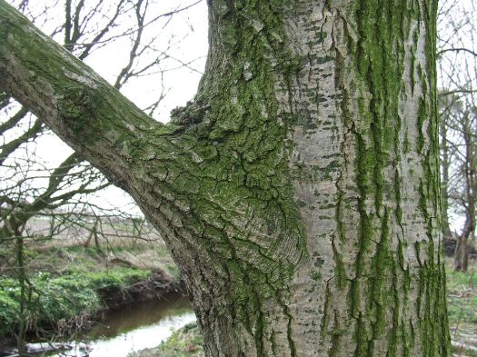
- Bark-Bark is the outermost layers of stems and roots of woody plants. Plants with bark include trees, woody vines, and shrubs. Bark refers to all the tissues outside the vascular cambium and is a nontechnical term. It overlays the wood and consists of the inner bark and the outer bark. The inner bark, which in older stems is living tissue, includes the innermost area of the periderm. The outer bark in older stems includes the dead tissue on the surface of the stems, along with parts of the innermost periderm and all the tissues on the outer side of the periderm. The outer bark on trees which lies external to the last formed periderm is also called the rhytidome
- Cork-Cork cambium (pl. cambia or cambiums) is a tissue found in many vascular plants as part of the epidermis. The cork cambium is a lateral meristem and is responsible for secondary growth that replaces the epidermis in roots and stems. It is found in woody and many herbaceous dicots, gymnosperms and some monocots (monocots usually lack secondary growth). It is one of the plant’s meristems – the series of tissues consisting of embryonic (incompletely differentiated) cells from which the plant grows. It is one of the many layers of bark, between the cork and primary phloem. The function of cork cambium is to produce the cork, a tough protective material.
- Phloem-In vascular plants, phloem is the living tissue that transports the soluble organic compounds made during photosynthesis and known as photosynthates, in particular the sugar sucrose, to parts of the plant where needed. This transport process is called translocation. In trees, the phloem is the innermost layer of the bark, hence the name, derived from the Greek word φλοιός (phloios) meaning “bark”. The term was introduced by Nägeli in 1858
- Vascular cambium-The vascular cambium (also called main cambium, wood cambium, bifacial cambium; plural cambia) is a plant tissue located between the xylem and the phloem in the stems and roots of vascular plants. It is a cylinder of unspecialized meristem cells that divide to form secondary vascular tissues. It is the source of both secondary xylem growth inwards towards the pith, and secondary phloem growth outwards to the bark. Unlike the xylem and phloem, it does not transport water, minerals or food through the plant.
- Heartwood and sapwood-Heartwood is wood that as a result of a naturally occurring chemical transformation has become more resistant to decay. Heartwood formation is a genetically programmed process that occurs spontaneously. Some uncertainty exists as to whether the wood dies during heartwood formation, as it can still chemically react to decay organisms, but only once.
- Branch collar-A branch collar is the often visible swelling in a woody plant that forms at the base of a branch where it is attached to its parent branch or to the tree’s trunk. The top of the branch collar consists of dense interlocking wood grain,[1] which provides mechanical support to the branch attachment. Branch collars can also be flat or somewhat recessed into the trunk or parent branch, as in some conifers. The collar consists primarily of tissues connected directly to the tree’s main trunk or stem, not to the branch.
- Root anatomy
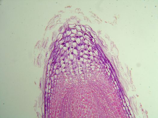
- Root structure-In vascular plants, the root is the organ of a plant that typically lies below the surface of the soil. Roots can also be aerial or aerating, that is, growing up above the ground or especially above water. Furthermore, a stem normally occurring below ground is not exceptional either (see rhizome). Therefore, the root is best defined as the non-leaf, non-nodes bearing parts of the plant’s body. However, important internal structural differences between stems and roots exist.
History
About 300 BC Theophrastus wrote a number of plant treatises, only two of which survive, Enquiry into Plants (Περὶ φυτῶν ἱστορία), and On the Causes of Plants (Περὶ φυτῶν αἰτιῶν). He developed concepts of plant morphology and classification, which did not withstand the scientific scrutiny of the Renaissance.
A Swiss physician and botanist, Gaspard Bauhin, introduced binomial nomenclature into plant taxonomy. He published Pinax theatri botanici in 1596, which was the first to use this convention for naming of species. His criteria for classification included natural relationships, or ‘affinities’, which in many cases were structural.
It was in the late 1600s that plant anatomy became refined into a modern science. Italian doctor and microscopist, Marcello Malpighi, was one of the two founders of plant anatomy. In 1671 he published his Anatomia Plantarum, the first major advance in plant physiogamy since Aristotle. The other founder was the British doctor Nehemiah Grew. He published An Idea of a Philosophical History of Plants in 1672 and The Anatomy of Plants in 1682. Grew is credited with the recognition of plant cells, although he called them ‘vesicles’ and ‘bladders’. He correctly identified and described the sexual organs of plants (flowers) and their parts.
In the eighteenth century, Carl Linnaeus established taxonomy based on structure, and his early work was with plant anatomy. While the exact structural level which is to be considered to be scientifically valid for comparison and differentiation has changed with the growth of knowledge, the basic principles were established by Linnaeus. He published his master work, Species Plantarum in 1753.
In 1802, French botanist Charles-François Brisseau de Mirbel, published Traité d’anatomie et de physiologie végétale (Treatise on Plant Anatomy and Physiology) establishing the beginnings of the science of plant cytology.
In 1812, Johann Jacob Paul Moldenhawer published Beyträge zur Anatomie der Pflanzen, describing microscopic studies of plant tissues.
In 1813 a Swiss botanist, Augustin Pyrame de Candolle, published Théorie élémentaire de la botanique, in which he argued that plant anatomy, not physiology, ought to be the sole basis for plant classification. Using a scientific basis, he established structural criteria for defining and separating plant genera.
In 1830, Franz Meyen published Phytotomie, the first comprehensive review of plant anatomy.
In 1838 German botanist Matthias Jakob Schleiden, published Contributions to Phytogenesis, stating, “the lower plants all consist of one cell, while the higher plants are composed of (many) individual cells” thus confirming and continuing Mirbel’s work.
A German-Polish botanist, Eduard Strasburger, described the mitotic process in plant cells and further demonstrated that new cell nuclei can only arise from the division of other pre-existing nuclei. His Studien über Protoplasma was published in 1876.
Gottlieb Haberlandt, a German botanist, studied plant physiology and classified plant tissue based upon function. On this basis, in 1884 he published Physiologische Pflanzenanatomie (Physiological Plant Anatomy) in which he described twelve types of tissue systems (absorptive, mechanical, photosynthetic, etc.).
British paleobotanists Dunkinfield Henry Scott and William Crawford Williamson described the structures of fossilized plants at the end of the nineteenth century. Scott’s Studies in Fossil Botany was published in 1900.
Following Charles Darwin‘s Origin of Species a Canadian botanist, Edward Charles Jeffrey, who was studying the comparative anatomy and phylogeny of different vascular plant groups, applied the theory to plants using the form and structure of plants to establish a number of evolutionary lines. He published his The Anatomy of Woody Plants in 1917.
The growth of comparative plant anatomy was spearheaded by British botanist Agnes Arber. She published Water Plants: A Study of Aquatic Angiosperms in 1920, Monocotyledons: A Morphological Study in 1925, and The Gramineae: A Study of Cereal, Bamboo and Grass in 1934.
Following World War II, Katherine Esau published, Plant Anatomy (1953), which became the definitive textbook on plant structure in North American universities and elsewhere, it was still in print as of 2006. She followed up with her Anatomy of seed plants in 1960.
